Imaging archives – international emergency medicine education project Lower extremity ultrasound dvt veins venous normal imaging anatomy findings Doppler ultrasound of lower limb arteries
Lower Extremity Arterial Ultrasound
Arterial lower extremity anatomy ultrasound vascular Ultrasound extremity arteries radiology Upper extremity arterial velocities ultrasound
Lower extremity arterial ultrasound
Arterial ultrasound of right upper extremity demonstrating no colorArterial sonography of the upper and lower extremities – sonographic [diagram] lower extremities diagramBilateral lower extremity arterial duplex.
Lower extremity arteries anatomyLower extremity arterial ultrasound Ultrasound assessment of lower extremity arteriesUltrasound assessment of lower extremity arteries.

Ultrasound vascular artery doppler arterial femoral peripheral duplex arteries waveform sonography nogu cfa imaging sfa pfa dopler evaluations carotid abdominal
Ultrasound extremity demonstrating arterial rtDoppler ultrasound limb arteries Ultrasound extremity arteriesFigure 1 from doppler ultrasonography of the lower extremity arteries.
Lower extremity arterial ultrasound worksheetDvt entire leg Lower extremity arterial ultrasoundUltrasound doppler limb arteries femoral popliteal vascular arnold.

Pdf doppler ultrasonography of the lower extremity arteries anatomy
Vascular ultrasound- lower extremity arterial anatomyDuplex lower arterial extremity bilateral study ultrasound vascular occlusion left case sfa disease radiology imaging Upper extremity arterial ultrasoundFigure 4 from doppler ultrasonography of the lower extremity arteries.
Perifer arteriell dubbelsidig scanning / vascular center / uc davisUltrasound lower extremity arteries Lower limb arterial anatomyUltrasound assessment of lower extremity arteries.

Ultrasound assessment of lower extremity arteries
Ultrasound assessment of lower extremity arteriesUltrasound dvt doppler vascular leg sonography arterial positioning peripheral epos limb diagnostic supine common procedure findings Dvt lower extremity anatomyLower extremity doppler arteries anatomy figure ultrasonography scanning guidelines.
Lower extremity arterial ultrasoundFigure 4 from doppler ultrasonography of the lower extremity arteries Ultrasound extremity arteries radiologyLower extremity doppler ultrasonography anatomy arteries figure scanning guidelines.
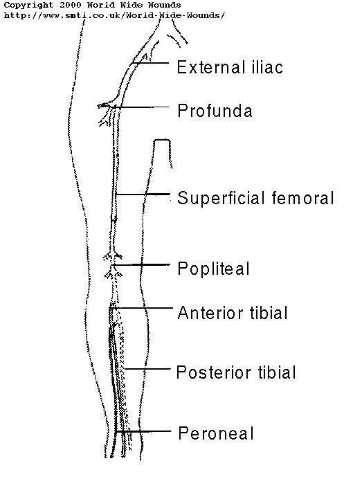
Extremity doppler anatomy arteries ultrasonography scanning guidelines
Pin von baloch auf vascularLower extremity arterial ultrasound Ultrasound extremity arteries radiologyPdf doppler ultrasonography of the lower extremity arteries anatomy.
Upper extremity arterial velocities ultrasoundUpper extremity arterial velocities ultrasound Peripheral arterial evaluationsLower extremity ultrasound disease peripheral artery arterial cardiovascular.

Doppler ultrasound of lower limb arteries
.
.


Upper Extremity Arterial Velocities Ultrasound

Arterial Sonography of the Upper and Lower Extremities – Sonographic

Arterial ultrasound of right upper extremity demonstrating no color

Figure 1 from Doppler ultrasonography of the lower extremity arteries

Perifer arteriell dubbelsidig Scanning / Vascular Center / Uc Davis

Ultrasound Assessment of Lower Extremity Arteries | Radiology Key
